Biology • Year 12 • Module 8 • Lesson 18
Hearing Loss, Cochlear Implants and Bone Conduction
Lock in the auditory pathway, the two categories of hearing loss, and the vocabulary needed to evaluate three hearing technologies.
1. Label the auditory pathway diagram
The diagram below shows the route from incoming sound to auditory perception. Write the missing labels into boxes A–H. Each label comes from the lesson’s Key Terms or Cards 1–2. 8 marks
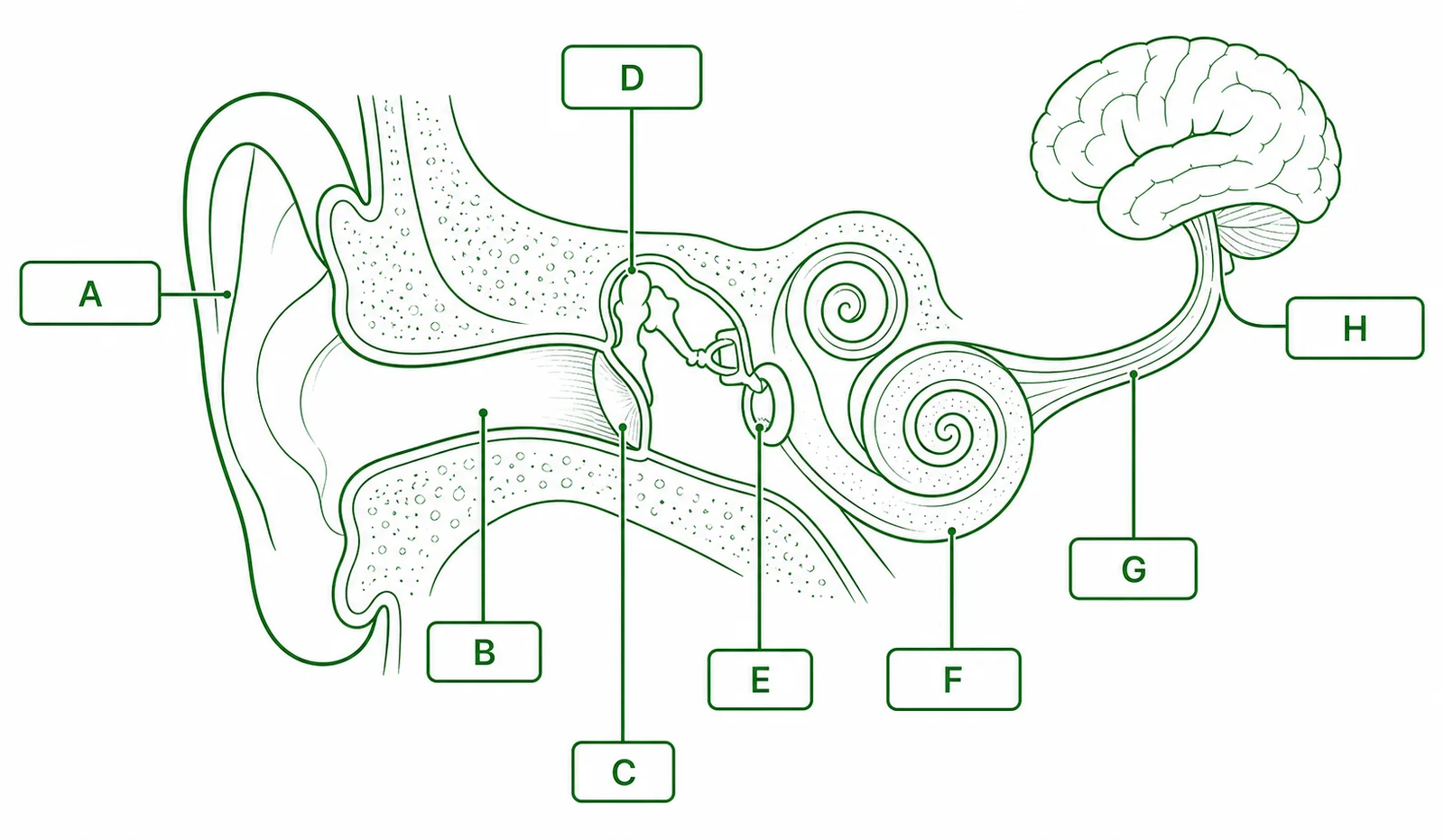
- A, outer ear structures (two structures) _______________________
- B, the three middle ear bones (collective name) _______________________
- C, fluid-filled coiled structure in the inner ear _______________________
- D, nerve carrying impulses from the cochlea to the brain _______________________
- E, lobe of the brain where sound is perceived _______________________
- F, function of the ossicles (one word) _______________________
- G, sensory cells inside the cochlea that transduce vibration _______________________
- H, property of the cochlea that encodes pitch along its length _______________________
| Box | Your label |
|---|---|
| A | |
| B | |
| C | |
| D | |
| E | |
| F | |
| G | |
| H |
2. Term–definition match
The ten definitions below are shuffled. In the right-hand column write the matching term from this list: cochlea, hair cells, tonotopic organisation, sensorineural hearing loss, conductive hearing loss, cochlear implant, bone conduction, osseointegration, auditory nerve, presbycusis. 10 marks
| # | Definition (shuffled) | Matching term |
|---|---|---|
| 2.1 | Mechanosensory cells in the organ of Corti whose stereocilia deflect in response to basilar membrane movement, opening ion channels and triggering neurotransmitter release. | |
| 2.2 | Hearing loss resulting from problems in the outer or middle ear that prevent sound waves from reaching the cochlea; the cochlea itself is intact and functional. | |
| 2.3 | The arrangement by which different frequencies are detected at different positions along the cochlear basilar membrane, high frequencies near the base, low frequencies near the apex. | |
| 2.4 | Cranial nerve VIII, which carries electrical impulses from the cochlea to the brainstem and auditory cortex. | |
| 2.5 | Age-related sensorineural hearing loss, typically affecting high-frequency sounds first. | |
| 2.6 | A surgically implanted device that delivers electrical pulses directly to the auditory nerve via an electrode array in the cochlea, bypassing non-functional hair cells. | |
| 2.7 | Hearing loss caused by damage to cochlear hair cells or the auditory nerve; cannot be fully corrected by amplification alone because the transducer is damaged. | |
| 2.8 | The fluid-filled, snail-shaped structure in the inner ear that converts mechanical vibration into electrical impulses. | |
| 2.9 | The transmission of sound vibrations directly through the skull bones to the cochlea, bypassing the outer and middle ear. | |
| 2.10 | The process by which living bone grows around and fuses with a titanium implant, anchoring it stably in the mastoid bone; used in BAHA devices. |
3. True or false, with correction
For each statement, circle T or F. If the statement is false, write the corrected version on the line below. 10 marks (1 T/F, 1 correction where needed)
3.1 A cochlear implant restores hearing to the same quality as natural hearing by replacing damaged hair cells with electronic equivalents. T / F
3.2 BAHA (bone-anchored hearing aid) is suitable for patients with conductive hearing loss because it transmits vibrations through the skull directly to a functional cochlea. T / F
3.3 In sensorineural hearing loss, the cochlear hair cells are intact, but the ossicles fail to amplify sound correctly. T / F
3.4 Hair cells in the mammalian cochlea do not regenerate after damage, so sensorineural hearing loss is typically permanent. T / F
3.5 A conventional hearing aid is the most appropriate technology for a patient with profound sensorineural hearing loss and no residual hair cell function. T / F
4. Function recall
Answer each in 1–2 sentences using precise terms from the lesson. 8 marks (2 each)
4.1 What is the function of the ossicles in the middle ear?
4.2 What is the function of the electrode array in a cochlear implant?
4.3 What is the function of tonotopic organisation in the cochlea?
4.4 What is the function of the transmitter coil in a cochlear implant system?
5. Fill the gaps, technology matching
Complete the paragraph using the word bank. Each word is used once. 8 marks
Hearing aids work by electronically amplifying sound so that residual cochlear _______________ can respond to a louder signal. They are most useful for mild-to-moderate _______________ hearing loss and for _______________ hearing loss where the cochlea is intact. For _______________ sensorineural hearing loss, a cochlear implant is indicated: its electrode array bypasses non-functional hair cells and directly stimulates the _______________. The implant’s external processor _______________ incoming sound into coded electrical signals. A bone-anchored hearing aid (BAHA) uses _______________ to deliver vibrations through the skull to the _______________, so it is only suitable when that structure is intact.
Q1, Auditory pathway labels
A: pinna and external auditory canal (either or both accepted). B: ossicles (malleus, incus, stapes). C: cochlea. D: auditory nerve (cranial nerve VIII). E: temporal lobe (auditory cortex). F: amplification / impedance matching. G: hair cells. H: tonotopic organisation.
Q2, Term–definition matches
2.1 hair cells • 2.2 conductive hearing loss • 2.3 tonotopic organisation • 2.4 auditory nerve • 2.5 presbycusis • 2.6 cochlear implant • 2.7 sensorineural hearing loss • 2.8 cochlea • 2.9 bone conduction • 2.10 osseointegration.
Q3, True / false with corrections
3.1 False. Correction: a cochlear implant does not restore normal hearing, it provides a different electrical signal through 12–22 electrode channels (compared with ~3,500 hair cell positions), which sounds different and requires extensive auditory rehabilitation. It provides access to sound, not natural hearing.
3.2 True.
3.3 False. Correction: in sensorineural hearing loss, the cochlear hair cells themselves are damaged (not the ossicles). The problem lies in the inner ear, hair cells cannot transduce basilar membrane vibration into electrical signals.
3.4 True.
3.5 False. Correction: for a patient with profound sensorineural hearing loss and no residual hair cell function, a conventional hearing aid is ineffective (it amplifies sound, but if no functional hair cells exist to transduce the signal, amplification is useless). A cochlear implant is the appropriate technology.
Q4.1, Function of the ossicles
The ossicles (malleus, incus, stapes) transmit and amplify vibrations from the tympanic membrane to the oval window of the cochlea. They also perform impedance matching, converting low-pressure, large-amplitude air vibrations into high-pressure, small-amplitude fluid vibrations in the cochlea.
Q4.2, Function of the electrode array
The electrode array (12–22 electrodes) is threaded into the scala tympani of the cochlea. Each electrode delivers precisely timed electrical pulses to auditory nerve fibres at a specific tonotopic position, directly depolarising those fibres and generating action potentials that travel to the auditory cortex, bypassing non-functional hair cells entirely.
Q4.3, Function of tonotopic organisation
Tonotopic organisation means different frequencies cause maximum basilar membrane resonance at different positions along the cochlea (high frequencies near the base, low near the apex). This spatial arrangement allows the cochlea to discriminate pitch, the brain interprets which region of the cochlea is most active as a particular frequency (pitch).
Q4.4, Function of the transmitter coil
The external transmitter coil transmits coded electrical signals from the external sound processor to the internal receiver-stimulator by radiofrequency electromagnetic induction through intact skin, without any wires piercing the skin surface.
Q5, Cloze paragraph (in order)
hair cells • sensorineural • conductive • profound • auditory nerve • amplifies • bone conduction • cochlea.