Biology • Year 12 • Module 7 • Lesson 10
The Innate Immune System
Lock in the key vocabulary, the components of the innate immune system, and the step-by-step process of phagocytosis.
1. Label the innate immune components diagram
The diagram below shows the main cellular and soluble components of the innate immune system arranged around a central hub. Write the correct label for each letter A–G. Each label names a component from the lesson's component table or Key Terms. 7 marks
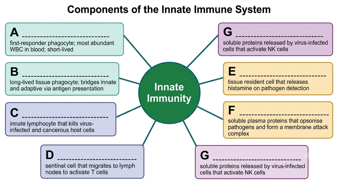
- A — first-responder phagocyte; most abundant WBC in blood; short-lived _______________________
- B — long-lived tissue phagocyte; bridges innate and adaptive via antigen presentation _______________________
- C — innate lymphocyte that kills virus-infected and cancerous host cells _______________________
- D — sentinel cell that migrates to lymph nodes to activate T cells _______________________
- E — tissue resident cell that releases histamine on pathogen detection _______________________
- F — soluble plasma proteins that opsonise pathogens and form a membrane attack complex _______________________
- G — soluble proteins released by virus-infected cells that activate NK cells and warn neighbours _______________________
| Box | Your label |
|---|---|
| A | |
| B | |
| C | |
| D | |
| E | |
| F | |
| G |
2. Term–definition match
The ten definitions below are shuffled. In the right-hand column write the matching term from this list: innate immunity, phagocytosis, phagocyte, pattern recognition receptor, natural killer cell, opsonisation, PAMP, phagosome, chemotaxis, phagolysosome. 10 marks
| # | Definition (shuffled) | Matching term |
|---|---|---|
| 2.1 | A fast, non-specific immune defence present from birth; responds to broad molecular patterns without prior exposure to the pathogen. | |
| 2.2 | The process by which immune cells engulf pathogens or debris by extending pseudopods to form an internal vesicle. | |
| 2.3 | A white blood cell such as a neutrophil or macrophage that performs the engulfing step of the process above. | |
| 2.4 | A receptor on immune cells that detects broad molecular patterns shared by many pathogens but not found on healthy host cells. | |
| 2.5 | An innate immune cell that kills host cells with reduced or absent MHC class I markers — including virus-infected and cancerous cells. | |
| 2.6 | The process of coating a pathogen with complement proteins or antibodies so that phagocytes can bind and engulf it more effectively. | |
| 2.7 | A pathogen-associated molecular pattern; a broad chemical signature found on pathogens (e.g. bacterial LPS) that is recognised by innate receptors. | |
| 2.8 | The membrane-bound vesicle formed inside a phagocyte after it engulfs a pathogen. | |
| 2.9 | Directed movement of a cell along a chemical concentration gradient — how phagocytes migrate toward a site of infection. | |
| 2.10 | The structure formed when a lysosome fuses with the vesicle containing the engulfed pathogen; the site of enzymatic digestion. |
3. True or false — with correction
For each statement, circle T or F. If the statement is false, write the corrected version on the line provided. 10 marks (1 for T/F, 1 for each correction)
3.1 Natural killer cells engulf and digest virus particles directly, the same way neutrophils destroy bacteria. T / F
3.2 The innate immune system has no memory — each exposure triggers the same response. T / F
3.3 Dendritic cells bridge the innate and adaptive immune systems by presenting antigens to T cells after engulfing pathogens. T / F
3.4 Mast cells release histamine when they detect pathogen-associated molecular patterns, causing vasodilation and increased capillary permeability. T / F
3.5 Innate immunity is weak and unimportant because it is non-specific — the adaptive immune system does all the real work. T / F
4. Function recall
Answer each in 1–2 sentences using precise terms from the lesson. 8 marks (2 each)
4.1 What is the function of pattern recognition receptors (PRRs) in the innate immune response?
4.2 What is the function of perforin in the NK cell killing mechanism?
4.3 What is the function of opsonisation in phagocytosis?
4.4 What is the function of MHC class I molecules on the surface of healthy host cells, in the context of NK cell surveillance?
5. Cloze — complete the phagocytosis paragraph
Select the correct word from the bank and write it in each blank. Each word is used once. 8 marks
Word bank: chemotaxis · phagolysosome · MHC II · pseudopods · PAMPs · lysosome · phagosome · T cells
When a neutrophil detects a bacterial infection, it follows a chemical concentration gradient by (5.1) _______________________ toward the site. The neutrophil's pattern recognition receptors bind (5.2) _______________________ on the pathogen surface — the adherence step. The cell then extends membrane projections called (5.3) _______________________ that wrap around the pathogen and close off behind it, forming an internal vesicle called a (5.4) _______________________. A (5.5) _______________________ then fuses with this vesicle, releasing digestive enzymes to form a (5.6) _______________________. After the pathogen is destroyed, antigen fragments are loaded onto (5.7) _______________________ molecules on the phagocyte's surface, where they are displayed for recognition by (5.8) _______________________ to initiate the adaptive immune response.
6. Innate vs adaptive — fill the comparison table
Complete the table below by writing the correct entry in each blank cell. 6 marks
| Feature | Innate immunity | Adaptive immunity |
|---|---|---|
| Speed of response | Minutes to hours | |
| Specificity | Highly specific — one antigen | |
| Immunological memory | No memory (classical view) | |
| Recognition method | Antigen receptors (BCR, TCR) | |
| Key cells | Neutrophils, macrophages, NK cells, mast cells | |
| Role in immune defence | Precision strike; creates memory |
Q1 — Innate components diagram labels
A: Neutrophils. B: Macrophages. C: Natural killer (NK) cells. D: Dendritic cells. E: Mast cells. F: Complement system. G: Interferons.
Q2 — Term–definition matches
2.1 innate immunity · 2.2 phagocytosis · 2.3 phagocyte · 2.4 pattern recognition receptor · 2.5 natural killer cell · 2.6 opsonisation · 2.7 PAMP · 2.8 phagosome · 2.9 chemotaxis · 2.10 phagolysosome.
Q3 — True / false with correction
3.1 False. Correction: NK cells do not engulf pathogens. They kill infected host cells by releasing perforin (which punches holes in the target cell membrane) and granzymes (which trigger apoptosis). Phagocytes such as neutrophils, not NK cells, engulf and digest pathogen particles.
3.2 True. (Accept: the classical definition applies for the HSC; note that recent research describes "trained innate immunity" in macrophages, but the HSC standard is no memory.)
3.3 True.
3.4 True.
3.5 False. Correction: The innate immune system is fast and powerful — it eliminates the majority of infections entirely on its own before the adaptive system is activated, and it shapes the adaptive response through cytokines and antigen presentation. People with innate immune deficiencies suffer life-threatening infections.
Q4.1 — Function of PRRs
PRRs detect broad molecular patterns called PAMPs (pathogen-associated molecular patterns) that are found on many pathogens but not on healthy host cells, allowing innate immune cells to rapidly identify and bind to a pathogen without prior exposure. This adherence step is essential for triggering phagocytosis.
Q4.2 — Function of perforin
Perforin is a pore-forming protein released by NK cells that punches holes in the target cell's plasma membrane. These pores allow granzymes (proteolytic enzymes) to enter the target cell and trigger apoptosis (programmed cell death), destroying the infected or cancerous host cell.
Q4.3 — Function of opsonisation
Opsonisation coats the surface of a pathogen with complement proteins or antibodies. Phagocytes have receptors that specifically recognise and bind to these coating molecules, making the adherence step dramatically faster and more effective — opsonised pathogens are far easier to phagocytose than uncoated ones.
Q4.4 — Function of MHC class I in NK surveillance
MHC class I molecules act as an "I am healthy" signal on normal host cells, inhibiting NK cells from killing them. When a cell is infected by a virus or becomes cancerous, MHC I expression is often reduced or lost; NK cells detect this "missing self" signal and destroy the target cell, eliminating the pathogen's hiding place.
Q5 — Cloze answers
5.1 chemotaxis · 5.2 PAMPs · 5.3 pseudopods · 5.4 phagosome · 5.5 lysosome · 5.6 phagolysosome · 5.7 MHC II · 5.8 T cells.
Q6 — Innate vs adaptive comparison table
Speed (adaptive): Days to weeks — must be activated. Specificity (innate): Non-specific — responds to broad PAMPs shared by many pathogens. Memory (adaptive): Immunological memory — faster, stronger response on re-exposure. Recognition (innate): Pattern recognition receptors (PRRs) detect PAMPs. Key cells (adaptive): B lymphocytes, T lymphocytes. Role (innate): First responder — contains infection and activates adaptive immunity.