Biology • Year 11 • Module 2 • Lesson 5
Cell Organisation, Review and Application
Apply the biological hierarchy to real data, real histology images, and an unfamiliar organ system, exactly the skills tested in HSC Section I and II.
1. Interpret SA:V ratio data, why being small or flat matters
The table below shows the surface area (SA), volume (V), and SA:V ratio for four model cells of different sizes and shapes. Use the data to answer the questions. 7 marks
| Cell model | Surface area (μm2) | Volume (μm3) | SA:V ratio |
|---|---|---|---|
| Cube, 2 μm side | 24 | 8 | 3.0 |
| Cube, 4 μm side | 96 | 64 | 1.5 |
| Cube, 8 μm side | 384 | 512 | 0.75 |
| Flat disc, 8 μm dia, 2 μm thick | ~154 | ~101 | ~1.53 |
1.1 Describe the trend in SA:V ratio as a cubic cell increases from 2 μm to 8 μm. 1 mark
1.2 Compare the SA:V ratio of the 8 μm cube to the flat disc of similar volume. Explain why the disc shape is advantageous for a cell that relies on simple diffusion across its surface. 3 marks
1.3 The human red blood cell is a biconcave disc approximately 8 μm in diameter. Using this data and your lesson knowledge, explain how the red blood cell’s shape is an example of the structure–function relationship. 3 marks
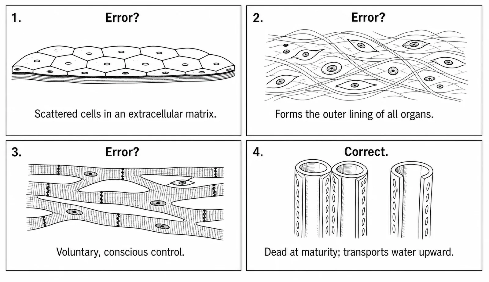
2. Histology diagram critique, spot the three errors
A student has produced the diagram below to show four tissue types and their key structural features. There are three biological errors in the diagram’s labels or descriptions. Identify each error and write the correction. 6 marks (2 per error: 1 identify, 1 correct)
2.1 Error 1: What is wrong?
Correction:
2.2 Error 2: What is wrong?
Correction:
2.3 Error 3: What is wrong?
Correction:
3. Cause-and-effect chain, from gene expression to organ function
The boxes on the left state a biological cause. For each one, complete the empty box on the right with the immediate biological effect. Use precise lesson terms. 5 marks
| Cause | → | Effect (complete this box) |
|---|---|---|
| A muscle cell gene is permanently switched on to produce large quantities of actin and myosin proteins. | → | |
| Thousands of differentiated cardiac muscle cells are connected by intercalated discs and contract simultaneously. | → | |
| Cardiac muscle tissue, epithelial tissue, connective tissue and nervous tissue are structurally integrated in the heart. | → | |
| The cardiovascular system connects the heart, arteries, capillaries and veins. | → | |
| All organ systems operate simultaneously under nervous and endocrine coordination. | → |
4. Apply the hierarchy to an unfamiliar organ system, the renal (urinary) system
You may not have studied the renal system in detail, but you can still apply the hierarchy. A nephron is the functional unit of the kidney. Each nephron contains a glomerulus (a knot of capillaries), a Bowman’s capsule (simple epithelium), a renal tubule (cuboidal epithelial cells with many mitochondria), and surrounding connective tissue. 6 marks
4.1 Identify the tissue types present in a nephron and state the function each performs in this context. 3 marks
4.2 Explain why the kidney qualifies as an organ using the definition from the lesson. 1 mark
4.3 What emergent property does the kidney possess that no single tissue type inside it could produce alone? 2 marks
Q1.1, SA:V trend (1 mark)
As the side length doubles (from 2 μm to 4 μm to 8 μm), the SA:V ratio halves each time (3.0 → 1.5 → 0.75). SA:V decreases as cell size increases because volume grows as the cube of the side length while surface area grows as the square.
Q1.2, Disc vs cube comparison (3 marks)
The 8 μm cube has SA:V of 0.75; the flat disc of similar volume has SA:V of ~1.53, roughly twice as high [1]. A higher SA:V means more surface membrane is available per unit of cytoplasm, so substances can diffuse in or out more rapidly relative to the volume of metabolically active material [1]. For a cell that relies on simple diffusion (no active transport), a higher SA:V ensures that O2, nutrients and waste products can be exchanged fast enough to meet the metabolic demands of the cell [1].
Q1.3, Red blood cell structure–function (3 marks)
The biconcave disc shape of the red blood cell is a direct example of the structure–function relationship [1]. The shape maximises SA:V compared to a sphere of the same volume, providing a large surface area for rapid diffusion of O2 into and CO2 out of the cell [1]. Additionally, the red blood cell has no nucleus and few organelles, maximising the volume available for haemoglobin, which carries O2every structural feature is optimised for the single function of gas transport [1].
Q2, Histology diagram errors (6 marks)
2.1 Error 1 (epithelial tissue label “scattered cells in extracellular matrix”): The label is describing connective tissue, not epithelial tissue. Correction: epithelial tissue is correctly described as a continuous sheet of tightly packed cells sitting on a basement membrane; it has little or no extracellular matrix between cells. [1 + 1]
2.2 Error 2 (connective tissue label “forms the outer lining of all organs”): Lining organs from the outside or inside is the function of epithelial tissue, not connective tissue. Correction: connective tissue supports, connects and protects other tissues; its cells are scattered in an extracellular matrix (e.g. collagen fibres); it does not form continuous linings. [1 + 1]
2.3 Error 3 (cardiac muscle labelled “voluntary, conscious control”): Cardiac muscle is involuntary, it is not under conscious control. Correction: cardiac muscle tissue is involuntary and self-stimulating (it generates its own electrical rhythm via the SA node / pacemaker); this distinguishes it from skeletal (voluntary, striated) muscle. [1 + 1]
Q3, Cause-and-effect chain (1 mark each)
Row 1: The cell becomes a specialised muscle cell, its structure is permanently modified to generate contractile force (e.g. high density of actin–myosin myofilaments).
Row 2: Cardiac muscle tissue generates enough pressure to drive blood through the heart, an emergent property impossible for any single cell.
Row 3: The heart becomes an organ capable of pumping blood in one direction, regulating its own rhythm and sustaining its own contractions, functions no single tissue can perform alone.
Row 4: The cardiovascular organ system can deliver O2 and nutrients to every cell in the body and remove CO2 and metabolic waste, whole-body circulation, impossible for the heart organ alone.
Row 5: The organism maintains homeostasis, stable internal conditions (e.g. body temperature, blood glucose, blood pH), an emergent property achievable only when all systems are simultaneously coordinated.
Q4.1, Tissue types in a nephron (3 marks)
Three tissue types are identifiable: (1) epithelial tissue, simple squamous epithelium lining the Bowman’s capsule (filtration surface) and cuboidal epithelium in the renal tubule (selective reabsorption and secretion) [1]; (2) connective tissue, surrounding and supporting the nephron components and providing the extracellular matrix scaffold [1]; (3) vascular/blood tissue within the glomerulus capillaries, delivering blood for filtration [1]. Accept “vascular tissue” or “blood” as a valid tissue type.
Q4.2, Kidney as an organ (1 mark)
The kidney qualifies as an organ because it contains two or more integrated tissue types (epithelial, connective, vascular/nervous) that together perform a complex, multi-step biological function, the filtration and selective reabsorption of blood to produce urine [1].
Q4.3, Emergent property of the kidney (2 marks)
The emergent property of the kidney is the ability to filter blood, selectively reabsorb useful substances (glucose, ions, water) and excrete metabolic wastes as urine [1]. No single tissue type can achieve this: epithelial tissue can filter or reabsorb, but it requires the vascular tissue to deliver blood under pressure and the connective tissue to maintain structural integrity, only the integrated organ performs the complete, regulated process [1].